As we enter the fourth year of the fastMOT project, we take a moment to reflect on our key achievements and milestones from the past 12 months. It has been a highly productive year, with significant progress across all work packages. Highlights also include six new publications and multiple presentations at leading international conferences.
All work packages continue to contribute to our shared vision: developing a revolutionary new light-sensing solution for non-invasive imaging of deep organ structures:
WP1: Detector development
In the current period, building on the previous fabrication of 10×10 SNSPD arrays on standard DBR substrates, we focused on the design and optimization of a new optical cavity stack. The goal was to achieve tailored absorption spectra with dual peaks at 785 nm and 1060 nm, enabling comparative studies in TD-NIRS, TD-DCS, and TD-SCOS. The stack design was optimized using a genetic algorithm and subsequently released for manufacturing. Initial optical characterization shows excellent agreement between measured and simulated absorption spectra. In parallel, NbTiN thin-film deposition is being carried out, and nanofabrication of the next SNSPD array generation is planned for the summer period, in line with the updated project plan.


WP2: Detector system development
We sucessfully installed the biggest individually addressed SNSPD system ever made by Single Quantum, the 10×10 system at Politecnico Milano. The detailed characterization of the system with the current version of the fastMOT chip was done at SQ labs, showing a detection efficiency > 50% at 1064 nm, an array average timing jitter of 70 ps, and an array average of 15 ns dead time. This detector system will be used for developing the optical tomograph for the project.
WP3: Physics light simulation and modelling
Collaborated with our partners in WP4 and WP5, we evaluated the performance of TD-NIRS, TD-DCS and TD-SCOS with the 1064nm Manny laser and the 10 by 10 SNSPD detectors, by using the Monte Carlo framework developed in Year 1. This investigation helps us identify the problem in the experimental phantom test and explore the benefits and challenges of using time gated approach in deep tissue monitoring. Further investigations will be implemented with realistic anatomical head geometry and different time gate position to evaluate the performance of this new system in functional deep brain imaging.
We are working on developing time-gated tomographic reconstruction algorithm for building 3D physiological parameter maps of DOT, DCT and SCOT, by using the time-gated approach compared with conventional reconstruction methods in optical tomography. We look forward to applying this new algorithm to the real measurements.
WP4: Tomographic work station
In WP4, we made significant progress toward assembling the tomograph prototype. We finalized the laser source configuration, and we designed all the other key sections of the system, including:
- The method for combining different light beams into a single delivery fiber
- The strategy for injecting light at multiple tissue positions to enable tomographic data acquisition
- The approach for focusing the collected light onto the new detector array composed of 100 SNSPDs
This detector array, together with its cryostat, helium compressor, 100-channel driver, and 120-channel time tagger, was delivered in December, marking the most important package received so far within the fastMOT project. All the other various pieces of the puzzle are arriving in the laboratory, and we are looking forward to connect everything together and carry out some experiments with this cutting-edge setup.
WP5: Laboratory and in vivo validation
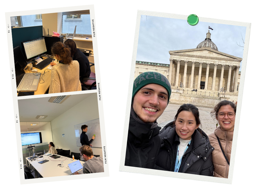
Work in WP5 this year has mainly been focused on preparing for upcoming measurements using the full 10×10 pixel system that will be fully operational in the laboratory at Politecnico di Milano (WP4) in the very near future. As part of this preparation, we have been collaborating with WP3 to define and run simulations to optimize measurements for deep-tissue sensitivity. An important part of this collaboration was the young researcher exchange where we spent three weeks working together in-person, exchanging know-how and ideas.
In addition, we have launched the fastMOT YouTube channel, where project partners share insights into our technology and ongoing work.
We look forward to an exciting fourth year, with upcoming events and continued innovation in medical imaging.
Subscribe to our newsletter and follow us on LinkedIn and YouTube to learn more about our work at fastMOT, and stay updated on our advances!
